Expanding the bandwidth of fluorescence-detected two-dimensional electronic spectroscopy using a broadband continuum probe pulse pair

Stephanie E. Sanders, Muyi Zhang, Ariba Javed and Jennifer P. Ogilvie

We demonstrate fluorescence-detected two-dimensional electronic spectroscopy (F-2DES) with a broadband, continuum probe pulse pair in the pump-probe geometry. The approach combines a pump pulse pair generated by an acousto-optic pulse-shaper with precise control of the relative pump pulse phase and time delay with a broadband, continuum probe pulse pair created using the Translating Wedge-based Identical pulses eNcoding System (TWINS). The continuum probe expands the spectral range of the detection axis and lengthens the waiting times that can be accessed in comparison to implementations of F-2DES using a single pulse-shaper. We employ phase-cycling of the pump pulse pair and take advantage of the separation of signals in the frequency domain to isolate rephasing and non-rephasing signals and optimize the signal-to-noise ratio. As proof of principle, we demonstrate broadband F-2DES on a laser dye and bacteriochlorophyll a.

Two-Dimensional Electronic Spectroscopy of the Far-Red-Light Photosystem II Reaction Center

Yogita Silori, Rhiannon Willow, Hoang H. Nguyen, Gaozhong Shen, Yin Song, Christopher J. Gisriel, Gary W. Brudvig, Donald A. Bryant, and Jennifer P. Ogilvie

Understanding the role of specific pigments in primary energy conversion in the photosystem II (PSII) reaction center has been impeded by the spectral overlap of its constituent pigments. When grown in far-red light, some cyanobacteria incorporate chlorophyll-f and chlorophyll-d into PSII, relieving the spectral congestion. We employ two- dimensional electronic spectroscopy to study PSII at 77 K from Synechococcus sp. PCC 7335 cells that were grown in far-red light (FRL-PSII). We observe the formation of a radical pair within ∼3 ps that we assign to ChlD1•−PD1•+. While PheoD1 is thought to act as the primary electron acceptor in PSII from cells grown in visible light, we see no evidence of its involvement, which we attribute to its reduction by dithionite treatment in our samples. Our work demonstrates that primary charge separation occurs between ChlD1 and PD1 in FRL-PSII, suggesting that PD1/PD2 may play an underappreciated role in PSII’s charge separation mechanism.

Charge Separation in the Photosystem II Reaction Center Resolved by Multispectral Two-Dimensional Electronic Spectroscopy

Hoang H. Nguyen, Yin Song, Elizabeth L. Maret, Yogita Silori, Rhiannon Willow, Charles F. Yocum, Jennifer P. Ogilvie

The photosystem II reaction center (PSII-RC) performs the primary energy conversion steps of oxygenic photosynthesis. While the PSII-RC has been studied extensively, the similar timescales of energy transfer and charge separation, and the severely overlapping pigment transitions in the Qy region have led to multiple models of its charge separation mechanism and excitonic structure. Here we combine two-dimensional electronic spectroscopy (2DES) with a continuum probe and two-dimensional electronic vibrational spectroscopy (2DEV) to study the cyt b559-D1D2 PSII-RC at 77K. This multispectral combination correlates the overlapping Qy excitons with distinct anion and pigment-specific Qx and mid-IR transitions to resolve the charge separation mechanism and excitonic structure. Through extensive simultaneous analysis of the multispectral 2D data we find that charge separation proceeds on multiple timescales from a highly delocalized excited state via a single pathway in which PheoD1 is the primary electron acceptor, while ChlD1 and PD1 act in concert as the primary electron donor.

Extracting the excitonic Hamiltonian of a chlorophyll dimer from broadband two-dimensional electronic spectroscopy

Kristina Zakutauskaite, Mindaugas Macernis , Hoang Nguyen , Jennifer P Ogilvie, and Darius Abramavicius

We apply Frenkel exciton theory to model the entire Q band of a tightly-bound chlorophyll dimer inspired by the photosynthetic reaction center of photosystem II. The potential of broadband two dimensional electronic spectroscopy experiment spanning the Qx and Qy regions to extract the parameters of the model dimer Hamiltonian is examined through theoretical simulations of the experiment. We find that the local nature of Qx excitation enables identification of molecular properties of the delocalized Qy excitons. Specifically we demonstrate that the cross-peak region, where excitation energy is resonant with Qy while detection is at Qx, contains specific spectral signatures that can reveal the full real-space molecular Hamiltonian, a task that is impossible by considering the Qy transitions alone. System-bath coupling and site energy disorder in realistic systems may limit the resolution of these spectral signatures due to spectral congestion.

Hidden Vibronic and Excitonic Structure and Vibronic Coherence Transfer in the Bacterial Reaction Center

Veronica R. Policht, Andrew Niedringhaus, Rhiannon Willow, Philip D. Laible, David F. Bocian, Christine Kirmaier, Dewey Holten, Tomáš Mančal, Jennifer P. Ogilvie

We report two-dimensional electronic spectroscopy (2DES) experiments on the bacterial reaction center (BRC) from purple bacteria, revealing hidden vibronic and excitonic structure. Through analysis of the coherent dynamics of the BRC, we identify multiple quasi-resonances between pigment vibrations and excitonic energy gaps, and vibronic coherence transfer processes that are typically neglected in standard models of photosynthetic energy transfer and charge separation. We support our assignment with control experiments on bacteriochlorophyll and simulations of the coherent dynamics using a reduced excitonic model of the BRC. We find that specific vibronic coherence processes can readily reveal weak exciton transitions. While the functional relevance of such processes is unclear, they provide a spectroscopic tool that uses vibrations as a window for observing excited state structure and dynamics elsewhere in the BRC via vibronic coupling. Vibronic coherence transfer reveals the upper exciton of the “special pair” that was weakly visible in previous 2DES experiments.

Phase-Modulated Rapid-Scanning Fluorescence-Detected Two-Dimensional Electronic Spectroscopy

Damianos Agathangelou, Ariba Javed, Francesco Sessa, Xavier Solinas, Manuel Joffre and Jennifer P. Ogilvie

Cover_v4.PNG

We present a rapid-scanning approach to fluorescence-detected two-dimensional electronic spectroscopy that combines acousto-optic phase-modulation with digital lock-in detection. The approach shifts the signal detection window to suppress 1/f laser noise and enables interferometric tracking of the time delays to allow for correction of spectral phase distortions and accurate phasing of the data. This use of digital lock-in detection enables acquisition of linear and nonlinear signals of interest in a single measurement. We demonstrate the method on a laser dye, measuring the linear fluorescence excitation spectrum, as well as rephasing, non-rephasing and absorptive fluorescence-detected two-dimensional electronic spectra.

Excitonic Structure and Charge Separation in the Heliobacterial Reaction Center Probed by Multispectral Multidimensional Spectroscopy

Yin Song, Riley Sechrist, Hoang Huy Nguyen, William Johnson, Darius Abramavičius, Kevin Redding, Jennifer P. Ogilvie

Graphic+abstract-final.jpg

Photochemical reaction centers are the engines that drive photosynthesis. The reaction center from heliobacteria (HbRC) has been proposed to most closely resemble the common ancestor of photosynthetic reaction centers, motivating a detailed understanding of its structure-function relationship. The recent elucidation of the HbRC crystal structure motivates advanced spectroscopic studies of its excitonic structure and charge separation mechanism. We perform multispectral two-dimensional electronic spectroscopy of the HbRC and corresponding numerical simulations, resolving the electronic structure and testing and refining recent excitonic models. Through extensive examination of the kinetic data by lifetime density analysis and global target analysis, we reveal that charge separation proceeds via a single pathway in which the distinct A0 chlorophyll a pigment is the primary electron acceptor. In addition, we find strong delocalization of the initial excited state and charge separation intermediate. Our findings have general implications for the understanding of photosynthetic charge separation mechanisms, and how they might be tuned to achieve different functional goals.

Mechanistic Study of Charge Separation in a Nonfullerene Organic Donor−Acceptor Blend Using Multispectral Multidimensional Spectroscopy

Yin Song, Xiao Liu, Yongxi Li, Hoang Huy Nguyen, Rong Duan, Kevin J. Kubarych, Stephen R. Forrest, and Jennifer P. Ogilvie*
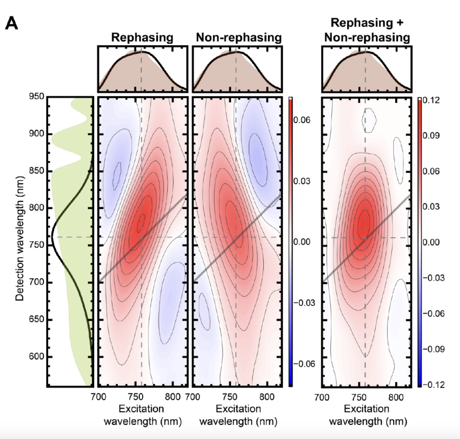
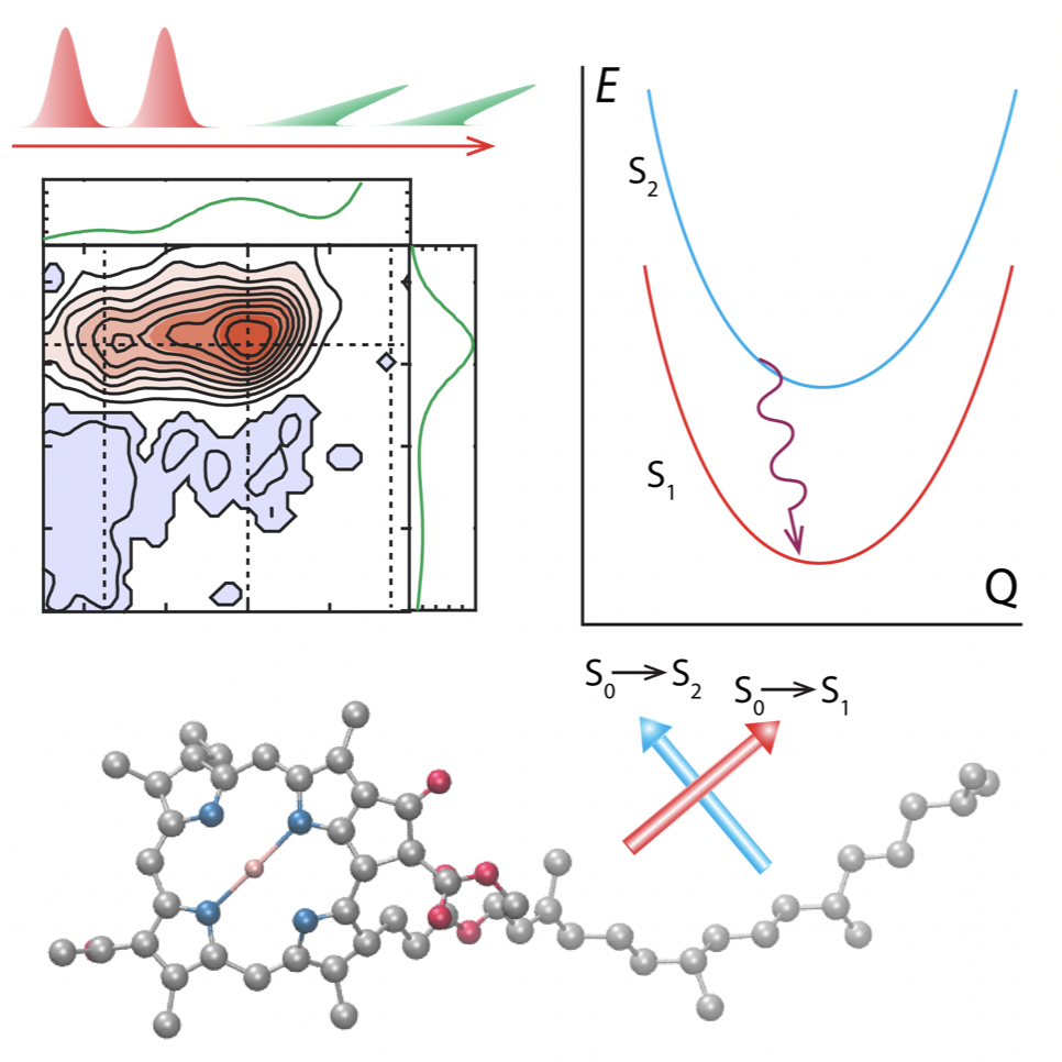
Organic photovoltaics (OPVs) based on nonfullerene acceptors are now approaching commercially viable efficiencies. One key to their success is efficient charge separation with low potential loss at the donor−acceptor heterojunction. Due to the lack of spectroscopic probes, open questions remain about the mechanisms of charge separation. Here, we study charge separation of a model system composed of the donor, poly[(2,6-(4,8-bis(5-(2-ethylhexyl)thiophen- 2-yl)-benzo[1,2-b:4,5-b′]dithiophene))-alt-(5,5-(1′,3′-di-2-thienyl-5′,7′-bis(2-ethylhexyl)benzo- [1′,2′-c:4′,5′-c′]dithiophene-4,8-dione) (PBDB-T), and the nonfullerene acceptor, 3,9-bis(2- methylene-(3-(1,1-dicyanomethylene)-indanone))-5,5,11,11-tetrakis(4-hexylphenyl)-dithieno[2,3- d:2′,3′-d′]-s-indaceno[1,2-b:5,6-b′]dithiophene (ITIC), using multidimensional spectroscopy spanning the visible to the mid-infrared. We find that bound polaron pairs (BPPs) generated within ITIC domains play a dominant role in efficient hole transfer, transitioning to delocalized polarons within 100 fs. The weak electron−hole binding within the BPPs and the resulting polaron delocalization are key factors for efficient charge separation at nearly zero driving force. Our work provides useful insight into how to further improve the power conversion efficiency in OPVs.

Two-dimensional electronic Stark spectroscopy of a photosynthetic dimer

Hoang H. Nguyen, Anton D. Loukianov, Jennifer P. Ogilvie and Darius Abramavicius

Screen Shot 2020-10-14 at 4.55.07 PM.png

Stark spectroscopy, which measures changes in the linear absorption of a sample in the presence of an external DC electric field, is a powerful experimental tool for probing the existence of charge-transfer (CT) states in photosynthetic systems. CT states often have small transition dipole moments, making them insensitive to other spectroscopic methods, but are particularly sensitive to Stark spectroscopy due to their large permanent dipole moment. In a previous study, we demonstrated a new experimental method, two- dimensional electronic Stark spectroscopy (2DESS), which combines two-dimensional electronic spectroscopy (2DES) and Stark spec- troscopy. In order to understand how the presence of CT states manifest in 2DESS, here, we perform computational modeling and calculations of 2DESS as well as 2DES and Stark spectra, studying a photosynthetic dimer inspired by the photosystem II reaction center. We identify specific cases where qualitatively different sets of system parameters produce similar Stark and 2DES spectra but significantly different 2DESS spectra, showing the potential for 2DESS to aid in identifying CT states and their coupling to excitonic states.

Observation of Ultrafast Coherence Transfer and Degenerate States with Polarization-Controlled Two-Dimensional Electronic Spectroscopy

Andy S. Sardjan, Floris P. Westerman, Jennifer P. Ogilvie, and Thomas L. C. Jansen

Screen Shot 2021-02-07 at 6.51.29 PM.png

Optical spectroscopy is a powerful tool to interrogate quantum states of matter. We present simulation results for 8 the cross-polarized two-dimensional electronic spectra of the light- 9 harvesting system LH2 of purple bacteria. We identify a spectral feature on the diagonal, which we assign to ultrafast coherence transfer between degenerate states. The implication for the interpretation of previous experiments on different systems and the potential use of this feature are discussed. In particular, we foresee that this kind of feature will be useful for identifying mixed degenerate states and for identifying the origin of symmetry breaking disorder in systems like LH2. Furthermore, this may help identify both vibrational and electronic states in biological systems such as proteins and solid-state materials such as hybrid perovskites.

Exploiting chemistry and molecular systems for quantum information science

Michael R. Wasielewski, Malcolm D. E. Forbes, Natia L. Frank, Karol Kowalski, Gregory D. Scholes, Joel Yuen-Zhou, Marc A. Baldo, Danna E. Freedman, Randall H. Goldsmith, Theodore Goodson III, Martin L. Kirk, James K. McCusker, Jennifer P. Ogilvie, David A. Shultz, Stefan Stoll & K. Birgitta Whaley

Screen Shot 2020-08-09 at 3.47.55 PM.png

The power of chemistry to prepare new molecules and materials has driven the quest for new approaches to solve problems having global societal impact, such as in renewable energy, healthcare and information science. In the latter case, the intrinsic quantum nature of the electronic, nuclear and spin degrees of freedom in molecules offers intriguing new possibilities to advance the emerging field of quantum information science. In this Perspective, which resulted from discussions by the co-authors at a US Department of Energy workshop held in November 2018, we discuss how chemical systems and reactions can impact quantum computing, communication and sensing. Hierarchical molecular design and synthesis, from small molecules to supramolecular assemblies, combined with new spectroscopic probes of quantum coherence and theoretical modelling of complex systems, offer a broad range of possibilities to realize practical quantum information science applications.

Quantum biology revisited

Jianshu Cao, Richard J. Cogdell, David F. Coker, Hong-Guang Duan, Jürgen Hauer, Ulrich Kleinekathöfer, Thomas L. C. Jansen, Tomáš Mančal, R. J. Dwayne Miller, Jennifer P. Ogilvie, Valentyn I. Prokhorenko, Thomas Renger, Howe-Siang Tan, Roel Tempelaar, Michael Thorwart, Erling Thyrhaug, Sebastian Westenhoff and Donatas Zigmantas

Screen Shot 2020-08-09 at 6.27.08 PM.png

Photosynthesis is a highly optimized process from which valuable lessons can be learned about the operating principles in nature. Its primary steps involve energy transport operating near theoretical quantum limits in effi- ciency. Recently, extensive research was motivated by the hypothesis that nature used quantum coherences to direct energy transfer. This body of work, a cornerstone for the field of quantum biology, rests on the interpretation of small-amplitude oscillations in two-dimensional electronic spectra of photosynthetic complexes. This Review discusses recent work reexamining these claims and demonstrates that interexciton coherences are too short lived to have any functional significance in photosynthetic energy transfer. Instead, the observed long-lived coherences originate from impulsively excited vibrations, generally observed in femtosecond spectroscopy. These efforts, collectively, lead to a more detailed understanding of the quantum aspects of dissipation. Nature, rather than trying to avoid dissipation, exploits it via engineering of exciton-bath interaction to create efficient energy flow.

Efficient Charge Generation via Hole Transfer in Dilute Organic Donor–Fullerene Blends

Yin Song, Alexander Schubert, Xiao Liu, Srijana Bhandari, Stephen R. Forrest, Barry D. Dunietz, Eitan Geva, and Jennifer P. Ogilvie*

Cover art-v3-02.png

Efficient organic photovoltaics (OPVs) require broadband charge photogeneration with near-unity quantum yield. This can only be achieved by exploiting all pathways that generate charge. Electron transfer from organic donors to acceptors has been well-studied and is considered the primary path to charge photogeneration in OPVs. In contrast, much less is known about the hole transfer pathway. Here we study charge photogeneration in an archetypal system comprising tetraphenyldibenzoperiflanthene:C70 blends using our recently developed multispectral two-dimensional electronic spectroscopy (M-2DES), supported by time-dependent density functional theory and fully quantum-mechanical Fermi’s golden rule rate calculations. Our approach identifies in real time two rapid charge transfer pathways that are confirmed through computational analysis. Surprisingly, we find that both electron and hole transfer occur with comparable rates and efficiencies, facilitated by donor–acceptor electronic interactions. Our results highlight the importance of the hole transfer pathway for optimizing the efficiency of OPV devices employing small-molecule heterojunctions.

Vibronic structure of photosynthetic pigments probed by polarized two-dimensional electronic spectroscopy and ab initio calculations

Yin Song, Alexander Schubert, Elizabeth Maret, Ryan K. Burdick, Barry D. Dunietz, Eitan Geva and Jennifer P. Ogilvie

Screen Shot 2020-08-09 at 8.25.31 PM.png

Bacteriochlorophyll a (Bchla) and chlorophyll a (Chla) play important dual roles as light- absorbers in photosynthetic antennae and initiators of primary charge-separation in photosynthetic reaction centers. Questions remain about the interplay of electronic and vibrational states within the low energy Q-band and its effect on the photoexcited dynamics. Here we study Bchla and Chla using two-dimensional electronic spectroscopy and state-of-the-art theoretical calculations. The Q-band of Bchla is comprised of two spectrally-separated bands at an angle of 75°. The Q-band of Chla contains two electronic transitions close in energy that are likely mixed via vibronic coupling. The internal conversion rates of Bchla and Chla are found to be 11 ps-1 and 38-50 ps-1, respectively. Our comparative study of these primary pigments highlights the interplay between their electronic structure and resulting photoexcited dynamics and may facilitate a mechanistic understanding of energy conversion in the many natural and artificial photosynthetic systems employing these pigments.

Multispectral multidimensional spectrometer spanning the ultraviolet to the mid-infrared

Yin Song, Arkaprabha Konar, Riley Sechrist, Ved Prakash Roy, Rong Duan, Jared Dziurgot, Veronica Policht, Yassel Acosta Matutes, Kevin J. Kubarych, and Jennifer P. Ogilvie

Screen Shot 2020-08-09 at 8.15.29 PM.png

Multidimensional spectroscopy is the optical analog to nuclear magnetic resonance, probing dynamical processes with ultrafast time resolution. At optical frequencies, the technical challenges of multidimensional spectroscopy have hindered its progress until recently, where advances in laser sources and pulse-shaping have removed many obstacles to its implementation. Multidi- mensional spectroscopy in the visible and infrared (IR) regimes has already enabled respective advances in our understanding of photosynthesis and the structural rearrangements of liquid water. A frontier of ultrafast spectroscopy is to extend and combine multidimensional techniques and frequency ranges, which have been largely restricted to operating in the distinct visible or IR regimes. By employing two independent amplifiers seeded by a single oscillator, it is straightforward to span a wide range of time scales (femtoseconds to seconds), all of which are often relevant to the most important energy conversion and catalysis problems in chemistry, physics, and materials science. Complex condensed phase systems have optical transitions spanning the ultraviolet (UV) to the IR and exhibit dynamics relevant to function on time scales of femtoseconds to seconds and beyond. We describe the development of the Multispectral Multidimensional Nonlinear Spectrometer (MMDS) to enable studies of dynami- cal processes in atomic, molecular, and material systems spanning femtoseconds to seconds, from the UV to the IR regimes. The MMDS employs pulse-shaping methods to provide an easy-to-use instrument with an unprecedented spectral range that enables unique combination spectroscopies. We demonstrate the multispectral capabilities of the MMDS on several model systems.

Simulating Fluorescence-Detected Two-Dimensional Electronic Spectroscopy of Multichromophoric Systems (Copy)

Tenzin Kunsel, Vivek Tiwari, Yassel Acosta Matutes, Alastair T. Gardiner, Richard J. Cogdell,‡ Jennifer P. Ogilvie,§ and Thomas L. C. Jansen

Screen Shot 2021-01-21 at 1.50.18 PM.png

We present a theory for modeling fluorescence-detected two-dimensional electronic spectroscopy of multichromophoric systems. The theory is tested by comparison of the predicted spectra of the light-harvesting complex LH2 with experimental data. A qualitative explanation of the strong cross-peaks as compared to conventional two-dimensional electronic spectra is given. The strong cross-peaks are attributed to the clean ground-state signal that is revealed when the annihilation of exciton pairs created on the same LH2 complex cancels oppositely signed signals from the doubly excited state. This annihilation process occurs much faster than the nonradiative relaxation. Furthermore, the line shape difference is attributed to slow dynamics, exciton delocalization within the bands, and intraband exciton−exciton annihilation. This is in line with existing theories presented for model systems. We further propose the use of time-resolved fluorescence-detected two-dimensional spectroscopy to study state resolved exciton−exciton annihilation.

Characterization of Vibrational Coherence in Monomeric Bacteriochlorophyll a by Two-Dimensional Electronic Spectroscopy

Veronica R. Policht, Andrew Niedringhaus, and Jennifer P. Ogilvie

Screen Shot 2021-02-06 at 3.00.22 PM.png

Bacteriochlorophyll a (BChla) is the most abundant pigment found in the Bacterial Reaction Center (BRC) and light-harvesting proteins of photosynthetic purple and green bacteria. Recent two-dimensional electronic spectroscopy (2DES) studies of photosynthetic pigment−protein complexes including the BRC and the Fenna−Matthews− Olson (FMO) complex have shown oscillatory signals, or coherences, whose physical origin has been hotly debated. To better understand the observations of coherence in larger photosynthetic systems, it is important to carefully characterize the spectroscopic signatures of the monomeric pigments. Prior spectroscopic studies of BChla have differed significantly in their observations, with some studies reporting little to no coherence. Here we present evidence of strong coherences in monomeric BChla in isopropanol using 2DES at 77 K. We resolve many modes with frequencies that correspond well with known vibrational modes. We confirm their vibrational origin by comparing the 2D spectroscopic signatures with expectations based on a purely vibrational model.

Spatially-resolved fluorescence-detected two-dimensional electronic spectroscopy probes varying excitonic structure in photosynthetic bacteria

Vivek Tiwari, Yassel Acosta Matutes, Alastair T. Gardiner, Thomas L. C. Jansen, Richard J. Cogdell & Jennifer P. Ogilvie

Screen Shot 2020-08-09 at 8.12.24 PM.png

Conventional implementations of two-dimensional electronic spectroscopy typically spatially average over ~10^10 chromophores spread over ~10^4 micron square area, limiting their ability to characterize spatially heterogeneous samples. Here we present a variation of two-dimensional electronic spectroscopy that is capable of mapping spatially varying differences in excitonic structure, with sensitivity orders of magnitude better than conventional spatially-averaged electronic spectroscopies. The approach performs fluorescence-detection-based fully collinear two-dimensional electronic spectroscopy in a microscope, combining femtosecond time-resolution, sub-micron spatial resolution, and the sensitivity of fluorescence detection. We demonstrate the approach on a mixture of photosynthetic bacteria that are known to exhibit variations in electronic structure with growth conditions. Spatial variations in the constitution of mixed bacterial colonies manifests as spatially varying peak intensities in the measured two-dimensional contour maps, which exhibit distinct diagonal and cross-peaks that reflect differences in the excitonic structure of the bacterial proteins.

Electronic Interactions in the Bacterial Reaction Center Revealed by Two-Color 2D Electronic Spectroscopy

Arkaprabha Konar, Riley Sechrist, Yin Song, Veronica R. Policht, Philip D. Laible, David F. Bocian, Dewey Holten, Christine Kirmaier, and Jennifer P. Ogilvie

Screen%2BShot%2B2020-08-09%2Bat%2B8.30.27%2BPM.jpg

The bacterial reaction center (BRC) serves as an important model system for understanding the charge separation processes in photosynthesis. Knowledge of the electronic structure of the BRC is critical for understanding its charge separation mechanism. While it is well-accepted that the “special pair” pigments are strongly coupled, the degree of coupling among other BRC pigments has been thought to be relatively weak. Here we study the W(M250)V mutant BRC by two-color two-dimensional electronic spectroscopy to correlate changes in the Qx region with excitation of the Qy transitions. The resulting Qy–Qx cross-peaks provide a sensitive measure of the electronic interactions throughout the BRC pigment network and complement one-color 2D studies in which such interactions are often obscured by energy transfer and excited-state absorption signals. Our observations should motivate the refinement of electronic structure models of the BRC to facilitate improved understanding of the charge separation mechanism.

Strongly coupled bacteriochlorin dyad studied using phase-modulated fluorescence-detected two-dimensional electronic spectroscopy

Vivek Tiwari, Yassel Acosta Matutes, Akraprabha Konar, Zhanqian Yu, Marcin Ptaszek, David F. Bocian, Dewey Holten, Christine Kirmaier and Jennifer P. Ogilvie

Screen Shot 2021-01-30 at 5.39.30 PM.png

Fluorescence-detected two-dimensional electronic spectroscopy (F-2DES) projects the third-order non-linear polarization in a system as an excited electronic state population which is incoherently detected as fluorescence. Multiple variants of F-2DES have been developed. Here, we report phase-modulated F-2DES measurements on a strongly coupled symmetric bacteriochlorin dyad, a relevant ‘toy’ model for photosynthetic energy and charge transfer. Coherence map analysis shows that the strongest frequency observed in the dyad is well-separated from the excited state electronic energy gap, and is consistent with a vibrational frequency readily observed in bacteriochlorin monomers. Kinetic rate maps show a picosecond relaxation timescale between the excited states of the dyad. To our knowledge this is the first demonstration of coherence and kinetic analysis using the phase-modulation approach to F-2DES.